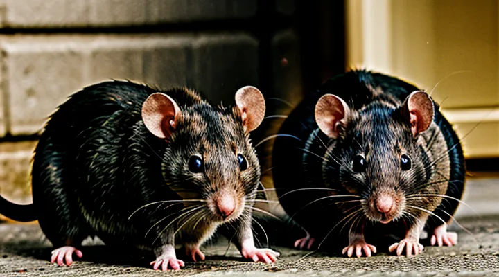
The Unique Structure of Rat Teeth
Enamel Composition and Iron
Rats possess continuously growing incisors whose outer layer is enamel enriched with iron compounds. Unlike the hydroxyapatite‑dominated enamel of most mammals, rat enamel incorporates ferric oxide particles that are deposited during tooth development. These iron deposits give the enamel a characteristic yellow‑brown hue, which persists despite the animal’s diet and environment.
The enamel structure consists of:
- A mineral matrix primarily composed of hydroxyapatite crystals.
- An iron‑rich layer situated just beneath the enamel surface.
- A thin outer enamel sheath where iron oxides are most concentrated.
During odontogenesis, iron ions are transported into the ameloblasts that lay down enamel. The ions bind to the forming crystal lattice, forming ferric oxide inclusions that alter the optical properties of the tissue. The resulting pigment absorbs shorter wavelengths of light, producing the visible yellow coloration.
The presence of iron also enhances enamel hardness and resistance to wear. Comparative studies show that rat incisors exhibit higher fracture toughness than enamel lacking iron, supporting the functional advantage of this adaptation for gnawing.
Dentin and Pulp
Rats’ incisors display a yellow hue because the outer enamel layer is thin and translucent, allowing the underlying dentin to dominate the visible color. Dentin, composed primarily of mineralized collagen and hydroxyapatite, possesses a natural amber tone. In rodents, dentin is continuously deposited as the tooth grows, maintaining the characteristic shade throughout the animal’s life.
The pulp chamber, situated centrally within the tooth, contains vascularized connective tissue, nerves, and odontoblasts that produce dentin. Although the pulp itself is not visible, its metabolic activity influences dentin formation. Rapid dentinogenesis ensures that newly formed dentin replaces worn enamel, preserving the yellow appearance.
Key factors linking dentin and pulp to tooth coloration:
- Translucent enamel: thin layer permits dentin’s pigment to show.
- Continuous dentin deposition: maintains amber color despite wear.
- Pulp vitality: supplies nutrients and signals for odontoblast function, regulating dentin quality.
Thus, the combination of a thin enamel cover, inherently pigmented dentin, and an active pulp system explains the persistent yellow coloration of rat teeth.
The Biological Reason for Yellow Teeth
Iron Pigmentation in Enamel
Rats display a distinctive yellow hue on their incisors because iron compounds become incorporated into the outer enamel layer during development. The process begins when iron ions from the diet are transported into ameloblasts, the cells responsible for enamel formation. Within these cells, iron binds to protein matrices and precipitates as ferric oxide particles that are subsequently deposited onto the enamel surface.
Key characteristics of iron‑based pigmentation:
- Ferric oxide crystals embed in the enamel’s outermost prisms, creating a stable, pigmented layer.
- The pigment resists abrasion, preserving the yellow coloration despite constant gnawing.
- Iron deposition occurs primarily during the maturation stage of enamel, when mineralization is most active.
The presence of iron alters the optical properties of enamel. Ferric particles absorb shorter wavelengths of visible light, shifting reflected light toward the yellow–orange spectrum. This effect is independent of bacterial plaque or dietary stains, which may add superficial discoloration but do not produce the intrinsic hue observed in healthy rodents.
Understanding iron pigmentation clarifies why rat incisors retain their characteristic color throughout life and differentiates this natural phenomenon from pathological discoloration seen in other mammals.
Constant Growth and Wear
Rats possess incisors that never cease to elongate because the teeth lack a closed root. An active growth zone at the base pushes the crown outward continuously. The outer surface consists of thin enamel, while the bulk of the tooth is made of dentin, a tissue naturally yellow in color. As the tooth lengthens, the newly formed dentin remains exposed until it is removed by chewing.
Gnawing provides the necessary abrasion to keep the incisors at a functional length. Each bite shaves off a thin layer of dentin, revealing fresh enamel and preventing over‑growth. When the rate of wear matches the rate of eruption, the teeth appear clean and relatively light. If wear is insufficient, excess dentin accumulates, increasing the visible yellow hue.
Factors that modify the balance between growth and wear include:
- Hardness of food items (seeds, nuts, wood) that increase abrasive action.
- Frequency of gnawing behavior, which directly determines material removal.
- Saliva composition, which softens dentin and may enhance staining from pigments in the diet.
- Environmental exposure to substances such as metal oxides that can deposit on the tooth surface.
The observed yellow coloration results from the constant interplay of relentless incisor growth and the steady removal of dentin through daily gnawing. When this equilibrium is maintained, rats keep functional, slightly yellow teeth; disruption of the balance leads to either overly long, darker incisors or excessive wear that reveals more enamel.
Dietary Influence on Tooth Color
Rats’ incisors are continuously growing and require constant wear to maintain proper length and enamel integrity. The color of these teeth reflects the composition of the diet, which can introduce pigments, alter mineral balance, and affect enamel maturation.
Pigments from food sources deposit on the enamel surface. Dark-colored grains, beetroot, carrots, and certain fruits contain carotenoids and anthocyanins that can stain the outer enamel layer. When rats consume these items regularly, the pigments accumulate, giving the teeth a yellowish hue.
Mineral imbalances influence enamel opacity. Diets low in calcium and phosphorus reduce the mineralization of the enamel, making it more translucent and allowing the underlying dentin, which is naturally yellow, to become visible. Conversely, excessive iron or manganese can produce a brownish tint that may be perceived as yellow when mixed with natural dentin color.
High‑sugar and high‑starch feeds promote bacterial fermentation in the oral cavity. Acidic by‑products erode the enamel surface, exposing dentin and accelerating discoloration. Fermentable carbohydrates also encourage the growth of pigmented oral microbes that contribute to tooth staining.
Typical dietary factors affecting rat tooth color:
- Carotenoid‑rich vegetables (e.g., carrots, pumpkin) – surface staining.
- Dark fruits and berries – anthocyanin deposition.
- Low‑calcium, low‑phosphorus diets – reduced enamel density.
- Excess iron or manganese supplements – metallic discoloration.
- High‑sugar, high‑starch foods – enamel erosion and dentin exposure.
- Fermentable fibers that support pigmented bacterial colonies – microbial staining.
Adjusting the diet to include balanced minerals, reducing pigmented foods, and limiting fermentable carbohydrates can maintain a lighter, healthier tooth appearance. Regular monitoring of dietary composition remains essential for preventing undesired coloration in rodent incisors.
Health Implications and Variations
Normal Versus Abnormal Discoloration
Rats’ incisors appear yellow due to the natural coloration of enamel and the presence of iron‑rich pigments. This hue is typical for healthy rodents and does not indicate disease. The yellow shade results from the combination of dentin, which is inherently darker, and a thin layer of enamel that allows the underlying color to show through.
Abnormal discoloration deviates from the expected yellow‑brown tone. Indicators include:
- Uniform black or gray staining, suggesting heavy metal accumulation or necrotic tissue.
- Red or pink hues, reflecting inflammation, infection, or hemorrhage within the pulp.
- Sudden loss of color, revealing translucency that may signal enamel erosion or nutritional deficiency.
Distinguishing normal from pathological coloration requires visual assessment of shade consistency, symmetry between upper and lower incisors, and the presence of accompanying signs such as excessive drooling, weight loss, or changes in chewing behavior. Consistent yellow coloration without ancillary symptoms confirms a normal condition; any deviation warrants veterinary examination.
Impact of Diet on Tooth Health
Rats develop yellow incisors when dietary composition interferes with the normal wear‑and‑tear of enamel. High‑starch or sugary foods promote bacterial growth that generates acid, which demineralizes the outer enamel layer. Continuous exposure to acidic by‑products accelerates discoloration and weakens the tooth surface.
Proteins and calcium are essential for maintaining dentin integrity. Diets lacking these nutrients reduce the mineral density of the incisors, allowing the underlying yellow dentin to become visible through the thinner enamel. Conversely, excessive mineral supplementation can lead to hyper‑mineralization, altering the translucency of the enamel and contributing to a yellow hue.
Key dietary factors affecting rat tooth health:
- Refined carbohydrates (e.g., cornmeal, sucrose) – increase acid production.
- Low calcium/phosphorus intake – diminish enamel strength.
- High fat content – reduces mastication efficiency, decreasing natural wear.
- Presence of abrasive fibers (e.g., coarse wood, hay) – promotes regular grinding, preserving enamel brightness.
- Vitamin C deficiency – impairs collagen formation in dentin, exposing yellow tissue.
Managing these variables through balanced feed formulations preserves the natural white appearance of rat incisors and prevents premature discoloration.
Signs of Dental Problems in Rats
Rats’ incisors grow throughout life, requiring constant wear from chewing. Yellow discoloration often signals that the natural grinding process is impaired, leading to dental complications that can affect health and longevity.
Signs of dental problems in rats include:
- Overgrown incisors that protrude beyond the lips or curl outward
- Misaligned or uneven teeth that create gaps or cause the mouth to appear asymmetrical
- Visible cracks, chips, or frayed edges on the enamel
- Persistent drooling or foamy saliva, especially after meals
- Reluctance to chew hard foods, resulting in a diet limited to soft items
- Weight loss despite adequate food availability
- Facial swelling or pus accumulation near the jaw
- Changes in grooming behavior, such as neglecting fur or excessive scratching around the mouth
- Reduced activity levels or signs of pain when the animal is handled
Early detection of these symptoms allows prompt veterinary intervention, which typically involves trimming overgrown teeth, addressing infections, and adjusting the diet to promote proper tooth wear. Maintaining appropriate chew toys and a fiber‑rich diet reduces the risk of dental issues and helps keep the incisors at a healthy color and length.
Comparative Analysis with Other Animals
Differences in Rodent and Human Teeth
Rats and humans possess fundamentally different dental architectures, a fact that directly influences the appearance of rat incisors. Rodent incisors grow continuously, whereas human teeth develop a fixed size and cease growth after eruption.
- Enamel: rats have a thin enamel layer covering only the front surface of the incisor; humans have a thick, complete enamel coating on all surfaces.
- Dentin: rodent incisors contain a large proportion of dentin that is exposed at the worn tip, giving a yellowish hue; human teeth retain dentin beneath enamel, keeping the crown white.
- Growth pattern: rodent incisors erupt from the root at a rate of 0.1 mm per day, requiring constant abrasion; human teeth do not erupt after maturity, eliminating the need for self‑sharpening.
- Diet and wear: rats chew hard materials that wear away enamel and expose dentin; humans typically consume softer foods, preserving enamel integrity.
- Root structure: rodents possess a single root that extends deep into the jawbone; humans have multi‑rooted molars and premolars, providing different mechanical support.
These anatomical distinctions explain why rodent incisors appear yellow while human teeth remain white under normal conditions. The exposed dentin in rats, combined with limited enamel protection and constant wear, results in the characteristic coloration observed in laboratory and wild specimens.
Other Animals with Pigmented Teeth
Dental pigmentation appears in many vertebrates, indicating that discolored incisors are not exclusive to rodents. Species with naturally pigmented or stained teeth include:
- Beavers – orange‑brown incisors colored by iron‑rich enamel.
- Musk shrews – yellowish incisors resulting from dietary carotenoids.
- African elephants – ivory tusks that develop a brownish hue with age due to mineral deposition.
- Peregrine falcons – darkened beak edges caused by melanin accumulation.
- Stingrays – denticles with reddish pigmentation linked to hemoglobin breakdown.
- Guppies – tiny teeth that acquire a pink tint from blood vessel proximity.
Pigmentation mechanisms vary. Iron compounds embed within enamel, producing amber tones in beavers. Carotenoid ingestion stains the dentin of small mammals such as shrews. Melanin synthesis within the odontoblast layer yields darkened beak or tooth surfaces in birds of prey. Mineralization and oxidation of organic matter cause progressive darkening in large mammals’ tusks. In aquatic species, blood‑derived pigments infiltrate dental structures during growth.
Understanding these patterns clarifies that rodent incisor discoloration reflects a broader biological phenomenon rather than an isolated anomaly. Comparative data on enamel composition, diet, and genetic factors across taxa provide a framework for interpreting the specific causes of yellowed rat teeth.
Maintaining Rat Dental Health
Appropriate Diet and Chewing Opportunities
Rats’ incisors grow continuously; the outer enamel is transparent while the underlying dentin is naturally yellow. When the enamel wears unevenly or the dentin becomes exposed, the teeth appear discolored.
A diet that promotes regular abrasion and limits staining supports healthy tooth coloration. Recommended components include:
- Fresh leafy greens (e.g., kale, romaine) – high fiber, low pigment.
- Raw carrots and celery – crisp texture encourages gnawing.
- Small portions of whole‑grain pellets – balanced nutrition without excess sugars.
- Limited fruit – occasional treat, avoiding high‑acid varieties that can discolor enamel.
Providing consistent chewing opportunities ensures even enamel wear and prevents overgrowth that reveals dentin. Effective options are:
- Untreated hardwood blocks (apple, maple) – durable, natural material.
- Cardboard tubes or paper rolls – readily available, encourages prolonged gnawing.
- Safe chew toys made of compressed wheat or sisal – designed for rodent dentition.
Combining a fiber‑rich, low‑stain diet with frequent, appropriate gnawing maintains the natural appearance of rat incisors and reduces the risk of yellowing.
Recognizing and Addressing Dental Issues
Rats rely on continuously growing incisors; discoloration often signals underlying dental problems. Yellow enamel can indicate plaque buildup, malnutrition, or disease, and may precede more serious conditions such as malocclusion or infection.
Visible indicators
- Yellow or brown staining on the front edge of incisors
- Uneven tooth length, with one side longer than the other
- Cracks, chips, or exposed pulp tissue
- Excessive drooling, difficulty chewing, or weight loss
Diagnostic approach A thorough visual inspection by a qualified veterinarian is the first step. Palpation of the jaw assesses tooth alignment, while radiographic imaging reveals root health and hidden lesions. Laboratory analysis of oral swabs identifies bacterial overgrowth.
Therapeutic measures
- Adjust diet to include high‑fiber foods (e.g., fresh vegetables, chewable grains) that naturally wear down teeth.
- Provide safe chew objects (wood blocks, mineral rods) to promote regular abrasion.
- Perform manual trimming of overgrown incisors under anesthesia when needed.
- Apply topical antimicrobial agents or prescribe systemic antibiotics for infections.
- In severe cases, extract damaged teeth and replace with prosthetic devices if feasible.
Preventive strategy Implement routine oral examinations during quarterly health checks. Maintain a balanced diet rich in fiber and low in sugary treats. Keep the cage environment enriched with chewable materials to ensure continuous tooth wear. Early detection and prompt treatment reduce the risk of progressive dental deterioration and improve overall wellbeing.
