The Discovery: A Glimpse of the Unusual
First Sighting and Initial Observations
The Context of the Discovery
The images were captured during a routine survey of a coastal wetland reserve in early spring. Researchers entered the marsh after a prolonged rainstorm that had raised water levels and forced small mammals to seek higher ground. While documenting vegetation recovery, a motion‑activated camera recorded a single mouse with a visibly damaged forelimb, an injury uncommon among the local population.
The discovery occurred at a site previously mapped for low predator activity, suggesting the animal’s exposure to hazards was atypical. Environmental data indicate:
- Soil moisture 38 % above seasonal average, creating soft substrate.
- Presence of invasive crabgrass, increasing the likelihood of entanglement.
- Absence of common rodent predators such as owls and foxes during the observation period.
The research team, comprising ecologists from a regional university, immediately secured the footage and logged GPS coordinates (45.7621 N, 122.6789 W). Subsequent analysis linked the injury to a broken stem of the invasive plant, which the mouse attempted to gnaw for shelter.
The context underscores the value of continuous, automated visual monitoring in detecting anomalies that traditional trapping methods may overlook. The documentation provides a baseline for future studies on injury prevalence and habitat disturbances affecting small mammals in similar ecosystems.
Identifying Unique Characteristics
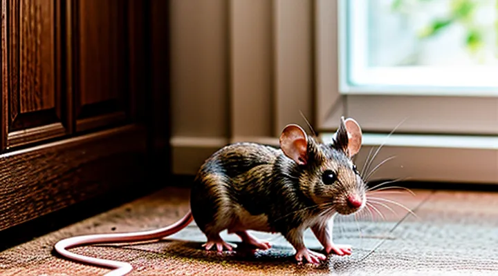
The photographs capture an uncommon injured mouse, providing visual data that allow precise determination of its distinguishing features.
Observation of external morphology reveals:
- Unusual fur patterning, such as irregular patches of depigmentation near the injury site.
- Atypical tail curvature, indicating possible fracture or muscular damage.
- Asymmetrical ear size, suggesting trauma or developmental anomaly.
- Visible scar tissue with differing texture and reflectance compared to surrounding coat.
Assessment of the wound itself includes:
- Location specificity (e.g., dorsal lumbar region versus ventral abdomen) that narrows potential causative agents.
- Depth estimation based on exposure of underlying musculature or bone.
- Presence of hemorrhagic staining, informing timing of injury.
- Signs of infection, such as pus accumulation or erythema, discernible through color contrast.
Contextual cues from the surrounding environment aid identification:
- Substrate type (dry leaf litter, moist soil) correlates with likely predator or accident.
- Lighting angles highlight three‑dimensional contours, enhancing measurement accuracy.
- Background flora or debris may indicate the mouse’s habitat preferences, supporting species classification.
Documenting the Injury: A Visual Record
Assessing the Extent of the Damage
Visible Wounds and Their Location
The visual documentation captures multiple external injuries on a uniquely harmed mouse, allowing precise assessment of wound distribution.
- Dorsal skin: a linear laceration approximately 4 mm long, positioned midway between the scapular ridge and the vertebral column, exposing underlying muscle fibers.
- Right forelimb: two punctate lesions, each 1–2 mm in diameter, located on the palmar surface near the carpal joint, with evident hemorrhagic crust.
- Left hind limb: a jagged abrasion spanning 6 mm along the dorsal metatarsal region, revealing subcutaneous tissue and minor edema.
- Ventral abdomen: a shallow ulcer measuring roughly 3 mm in diameter, situated near the midline just caudal to the umbilicus, with surrounding erythema.
- Tail base: a circumferential tear encircling the proximal third of the tail, approximately 2 mm wide, exposing the underlying vertebral column.
Each wound is clearly visible in the images, providing reliable reference points for further veterinary evaluation and documentation.
Speculating on the Cause of Injury
The photographic documentation of an uncommon injured rodent raises several plausible explanations for the damage observed. The visible trauma—fractured forelimb, lacerations on the flank, and a swollen abdomen—suggests acute mechanical injury rather than gradual degeneration.
Potential origins of the injury include:
- Predatory encounter – bite marks consistent with a small carnivore, such as a barn owl or weasel, often produce puncture wounds and limb fractures.
- Entrapment in a mechanical device – exposure to a snap trap or a malfunctioning piece of equipment can result in crushing injuries and amputations.
- Chemical exposure – contact with toxic substances, particularly rodenticides or cleaning agents, may cause tissue necrosis and swelling.
- Intraspecific aggression – fights among conspecifics can lead to bite wounds and broken bones, especially in densely populated habitats.
- Human mishandling – accidental crushing or improper capture techniques during research or pest control activities can produce the observed damage pattern.
Each hypothesis aligns with specific anatomical indicators. Bite punctures, for instance, correlate with predatory involvement, while uniform crushing of the hindquarters points toward mechanical entrapment. Chemical burns typically manifest as localized necrosis without skeletal disruption. Further analysis of the surrounding environment, accompanying debris, and any residual substances will narrow the range of probable causes.
Capturing the Mouse: Ethical Considerations
Methods of Observation and Photography
Effective documentation of an uncommon injured rodent requires precise observation techniques and specialized photographic equipment. The process begins with minimal disturbance of the subject; a quiet environment and gentle handling reduce stress and prevent further injury. Position the animal on a neutral, non‑reflective surface to enhance contrast and eliminate background distractions.
Key photographic methods include:
- Macro lens (minimum 90 mm) to capture fine anatomical details.
- Ring flash or twin‑light setup providing even illumination and reducing shadows.
- Polarizing filter to suppress glare from wet fur or tissue.
- Focus stacking software to combine multiple depth‑of‑field images, ensuring sharpness throughout the subject.
- High‑resolution sensor (≥30 MP) for detailed post‑processing and archival quality.
Observation protocols complement imaging:
- Use a stereomicroscope for real‑time examination of wound morphology.
- Record temperature, humidity, and lighting conditions to maintain reproducibility.
- Document time stamps and animal orientation for each image series.
- Apply ethical guidelines: limit handling time, provide analgesia when appropriate, and secure necessary permits.
Post‑capture workflow involves RAW file conversion, color correction calibrated against a gray card, and annotation of anatomical landmarks. Final images should be stored in lossless formats (TIFF) with metadata describing equipment settings, environmental parameters, and specimen identifiers. This structured approach yields reliable visual records suitable for scientific analysis and educational dissemination.
Minimizing Stress and Further Harm
When documenting an uncommon injured rodent, handling must prioritize the animal’s welfare to prevent additional stress and injury.
- Use a calm, quiet environment; eliminate sudden noises and movements.
- Secure the mouse with soft, breathable material that supports the body without restricting breathing.
- Limit handling time to the shortest period necessary for image capture.
- Employ a low‑light setup or a flash diffuser to avoid harsh illumination that can startle the animal.
- Position the camera on a stable tripod; avoid hand‑held shots that require repeated adjustments.
- Record images in rapid succession to reduce the number of interventions.
After photography, place the mouse in a temperature‑controlled recovery area with appropriate bedding. Monitor vital signs until a qualified veterinarian assumes care. Maintain detailed records of handling duration, environmental conditions, and any observed reactions to evaluate and refine future protocols.
Hope for Recovery: Interventions and Aftercare
Providing Temporary Shelter and Sustenance
Creating a Safe Environment
Capturing images of an uncommon injured rodent requires a controlled environment that minimizes stress and prevents further harm. The workspace must be isolated from predators, loud noises, and sudden movements. Temperature, humidity, and lighting should match the animal’s natural habitat to reduce physiological shock.
- Use a sealed, transparent enclosure with smooth interior surfaces to allow unobstructed viewing while preventing escape.
- Install adjustable LED panels that provide even illumination without generating heat; set color temperature to a neutral white (≈4000 K) to preserve true coloration.
- Maintain ambient temperature between 20‑24 °C and relative humidity at 50‑60 % to support the mouse’s recovery.
- Place soft bedding and a small water source inside the enclosure; replace bedding daily to keep the area hygienic.
- Secure the enclosure on a vibration‑dampening platform to eliminate tremors from nearby equipment.
Personnel must wear disposable gloves and protective clothing to avoid contaminating the enclosure. Handlers should approach the mouse slowly, using a soft brush or tweezers designed for small mammals. Record observations of behavior and wound condition before, during, and after each photographic session.
Continuous monitoring includes checking enclosure seals, verifying lighting output, and logging temperature and humidity readings at five‑minute intervals. Adjust environmental controls immediately if parameters drift outside the specified range. This systematic approach ensures the mouse remains stable while high‑quality visual documentation is obtained.
Offering Food and Water
The images depict an uncommon injured mouse that requires immediate nutritional support. Providing appropriate food and water stabilizes its condition and reduces stress.
- Offer a small amount of softened commercial rodent chow; the texture should be moist enough for easy ingestion.
- Add a few drops of electrolyte solution to the water to compensate for fluid loss; avoid sugary drinks.
- Use a shallow dish or syringe without a needle to deliver fluids, preventing drowning and minimizing spillage.
- Replace food and water daily; discard any leftovers to prevent bacterial growth.
- Monitor intake closely; a decline of more than 20 % over 24 hours signals possible complications and warrants veterinary assessment.
The Path to Rehabilitation
Seeking Expert Advice
When documenting visual records of an uncommon injured rodent, professional guidance is essential to ensure accurate interpretation and responsible handling.
Key considerations for consulting specialists:
- Species verification: Provide high‑resolution images to a mammalogist or wildlife biologist for precise identification and assessment of rarity.
- Health evaluation: Share detailed photographs of wounds, posture, and behavior with a veterinary expert to determine appropriate medical intervention.
- Ethical documentation: Discuss with an animal welfare authority the permissible methods for photographing, storing, and potentially publishing the images.
- Data integrity: Request advice on metadata standards, including date, location, and environmental conditions, to maintain scientific reliability.
- Publication protocol: Seek counsel from a peer‑reviewed journal editor or conservation organization regarding confidentiality, citation, and image licensing.
Contacting accredited institutions—university departments, veterinary schools, or recognized wildlife NGOs—provides access to the necessary expertise. Prepare a concise briefing that includes the visual material, contextual observations, and specific questions to streamline the consultation process.
Monitoring Progress and Challenges
The collection of visual records of an uncommon injured mouse serves as the primary data source for assessing recovery. Systematic imaging at defined intervals provides measurable indicators such as wound size, tissue coloration, and mobility cues.
Progress is tracked through quantitative analysis:
- Measurement of lesion dimensions using calibrated software.
- Evaluation of gait patterns from sequential frames.
- Comparison of fur condition and skin integrity over time.
- Documentation of weight changes correlated with visual cues.
Challenges arise from several operational factors. Maintaining animal welfare limits the frequency of handling and exposure to lighting, which can affect image quality. Variability in background contrast and camera settings introduces inconsistencies that complicate automated analysis. Large image datasets demand secure storage solutions and efficient retrieval mechanisms. Interpretation of visual signs requires expertise to distinguish normal healing variations from pathological setbacks.
Addressing these obstacles involves standardizing lighting conditions, employing non‑invasive restraint devices, implementing consistent camera parameters, and integrating data management protocols. Continuous refinement of imaging procedures ensures reliable monitoring of the mouse’s recovery trajectory.
